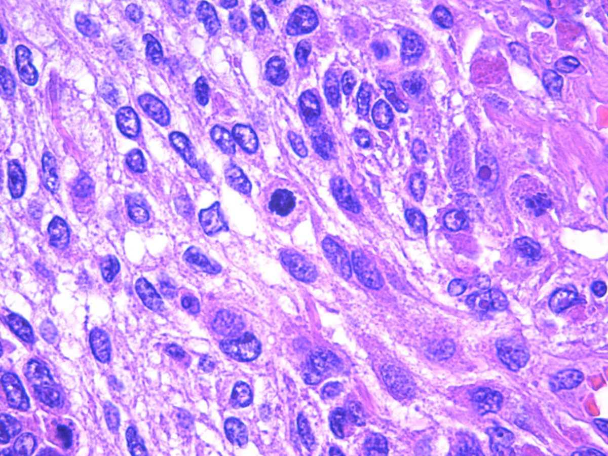

(k) metaphase II (l) anaphase II (m) telophase II (n) round spermatids (o) elongating spermatids (p) head of sper-matozoas. (a) polyploid nutri-tive cells with many heteropicnotic chromatin bodies (b) leptotene-zygotene (c) pachytene (d) diffuse stage (e) di-plotene (f) diakinesis (g) metaphase I (h, i) anaphase I (j) telophase I. You can see the photos of diplotene and diakinesis here : The only difference between this phase and Diakinesis is that The centrosomes reach the poles. ID characteristics that can help you recognize diplotene better: Answers without enough detail may be edited or deleted. Want to improve this post? Provide detailed answers to this question, including citations and an explanation of why your answer is correct. Here's an image which has nearly all the stages except telophase.Can someone label the stages?įinally am I correct? How do I identify them correctly? Could you site some authentic references? The sister chromatids of a dyad separate due to splitting of centromere.The dyads are arranged in circle or flower like fashion.One group is with 11 autosomes and the other group with 11 autosomes and a X chromosome.Two groups of chromosome are in the act of separation.Terminalisation of chiasmata completes.The chromosomes are aligned at the equitorial plate.Presence of twelve chromosomal elements (11 tetrad + X chromosome).The nucleus and nuclear membrane disappear completely.The bivalents appear rod, diamond or oval shaped.Terminalisation of chiasmata initiates.

Nucleolus and nuclear membrane begin to diappear.(can't be detected).The paired chromosomes separate from each other except for the chiasmata.The chromosomes are wooly in appearance.The chromosomes are thick and darkly stained.Crossing over between non sister chromatids of homologous chromosomes occur leading to formation of X shaped chiasmata.The sister chromatids separate to form tetrad a structure with four chromatids.The chromosomes show some degree of condensation.Nucleolus and nuclear membrane are present.(not visible).The single darkly stained X chromosome is present slightly off the periphery of the nucleus.Homologous chromosomes undergo synapsis forming bivalent.The uncondensed chromosomes are visible as a cloud of threads.Nuceolus and nuclear membrane are present.(not visible under compound.Late in this stage the chromosomes attach themselves by telomeres to the inner membrane of the nuclear envelope forming a bouquet.The single darkly stained X chromosome is found at the periphery of the nucleus.Each chromosome consist of two chromatids which are not visible.The uncondensed chromosomes are visible as a cloud of thin threads.

Note: The character in bold and italics is the identifying feature of the concerned stage. Specimen: Grasshopper ( Gesonula punctifrons) testis whose 2n= 23

Here's what I know and understand about meiotic stages: I need help so I decided to post this here.
#Prophase under microscope free#
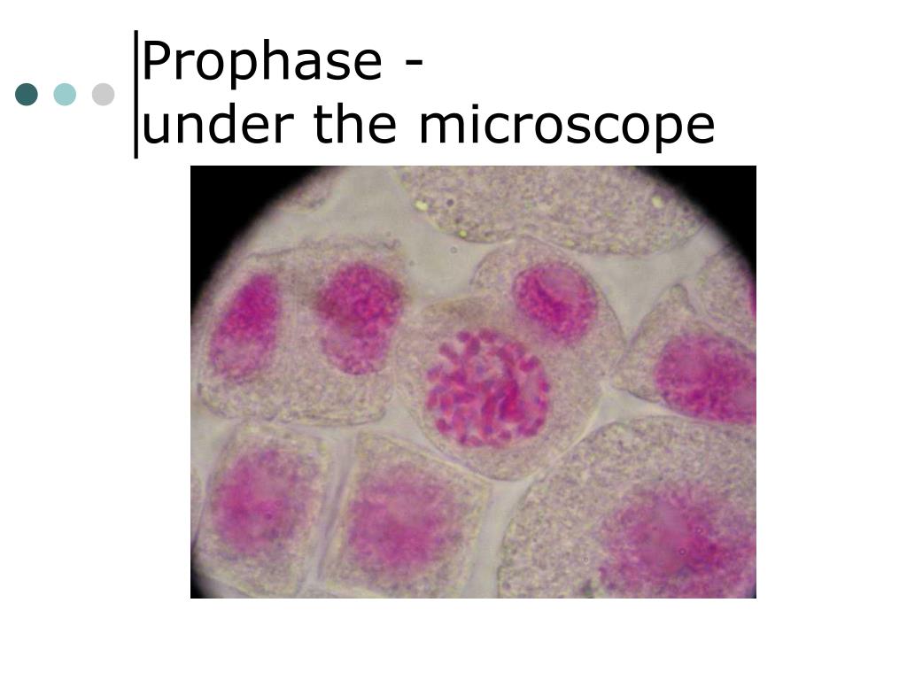
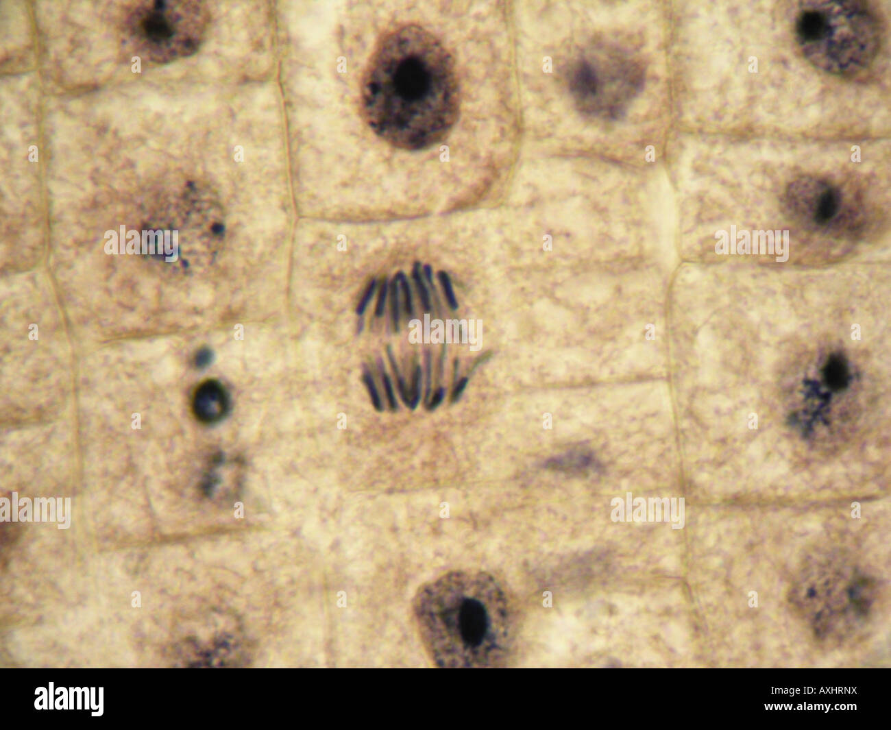
You will be looking at strands of DNA inside the cell! Note: The onion root tip slide is included free in your slide kit when you purchase a microscope from Microscope World.Ĭalled Prophase, the DNA molecules of theĬalled Metaphase, the chromosomes line up in the center of the cell, separate and become a pair of identical chromosomes.This is a question I'm troubled with as my exams are knocking at the door. If you have a microscope (400x) and a properly stained slide of the Onion root tip (or Allium root tip), you can see the phases in different cells, frozen in time. In the drawings below, you can see the chromosomes in the nucleus going through the process called mitosis, or division. One of these processes involves the splitting of the chromosomes. When a cell splits and becomes two, certain processes occur within the nucleus first. These are important for heredity and reproduction. For example, within the nucleus lie the chromosomes. There are various structures within the cell, but many are too difficult to see.


 0 kommentar(er)
